Addition and correction
Bio-milling technique for the size reduction of chemically synthesized BiMnO3 nanoplates
Baishakhi Mazumder, Imran Uddin, Shadab Khan, Venkat Ravi, Kaliaperumal Selvraj, Pankaj Poddar and Absar Ahmad
J. Mater. Chem., 2007, 17, 3910–3914 (DOI: 10.1039/b706154d). Amendment published 5th December 2007.
Recently, we reported a novel bio-milling technique where we showed that it was possible to reduce the size of the chemically synthesized particles into nanoparticles with size as small as 5 nm using a microbial process.1 In this article, we identified the particles synthesized by the wet-chemical method (synthesis procedure reported in a separate study) as perovskite BiMnO3.2 Our study was based on reacting the as-synthesized particles with fungus to demonstrate that over a period of time, fungus could actually break down the bigger particles into smaller ones, a mechanism which is common in nature.
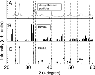
In this article, the phase identification of the material used as a model system for bio-milling—BiMnO3—was based on the previous reported work which originally reported the synthesis of the material.1,2 However after a careful analysis using the JCPDS database, it was later found that the phase reported by us in this article is probably BiOCl instead of BiMnO3. Other scientific details of the article remain unchanged (such as the bio-milling, protein analysis, etc.). Our previous structural analysis was due to the similarities in several XRD peaks of our data with the BiMnO3 as shown in the figure below.2,3 Texturing effects due to the plate-like particles were also thought to contribute in reduction of the peak intensities of some of the major peaks.1 The material BiOCl (bismuth oxide chloride) has tetragonal structure (space group: P4/nmm) with lattice constants: a = b = 3.89 Å and c = 7.37 Å. In the figure below, we have compared the XRD data of as-synthesized BiOCl nanoplates (panel A) with the BiMnO3 (panel B) and BiOCl XRD profiles as obtained from the JCPDS files 89-4544 and 85-0861 respectively. The lattice planes in the HRTEM images of the bio-milled nanoparticles (Fig. 6), reported in the article by Mazumder et al., were re-indexed to the lattice planes <212> and <113> corresponding to the lattice spacings of ~1.57 Å and 1.83 Å (±0.03 Å) respectively.1
Figure Comparison between the powder XRD profiles (A) reported in Mazumder et al., J. Mater. Chem., 2007, 17, 3910 for as synthesized particles; (B) reference peaks for the BiMnO3 phase taken from JCPDS file 89-4544; (C) reference peaks for the BiOCl phase taken from JCPDS file 85-0861.
References
1 B. Mazumder, I. Uddin, S. Khan, V. Ravi, K. Selvraj, P. Poddar and A. Ahmad, J. Mater. Chem., 2007, 17, 3910.
2 V. Samuel, S. C. Navale, A. D. Jadhav, A. B. Gaikwad and V. Ravi, Mater. Lett., 2007, 61, 1050.
3 H. Chiba, T. Atou, H. Faqir, M. Kikuchi, Y. Syono, Y. Murakami and D. Shindo, Solid State Ionics, 1998, 108, 193–199.
The Royal Society of Chemistry apologises for these errors and any consequent inconvenience to authors and readers.
Back to article