This Faraday discussion will be a hybrid event, allowing participation both in person and online.
Welcome
Join us in Sheffield, or online, in July 2022 for this addition to our Faraday Discussion series. For over 100 years and 300 meetings, Faraday Discussions have led the conversation on the study of the sciences lying between chemistry, physics and biology. Many of these Discussions have become landmark meetings in their field with the unique format allowing for in-depth discussions and opportunities to establish new collaborations.This Discussion is for established and early-career scientists, post-graduate students and industrial researchers interested in the expanding field of cryo-electron microscopy. Researchers in this area who based in cryo-EM facilities or research institutes, or in departments of biology, chemistry, structural biology, virology, cell biology, molecular biology or biophysics etc are all welcome. Oral and poster presentation opportunities are available to all these groups, and we invite you to submit an oral or poster abstract and join us to make your contribution alongside leaders in the field.
The Discussion will bring together expertise from the UK and the international cryo-electron microscopy community to discuss current developments and new challenges.
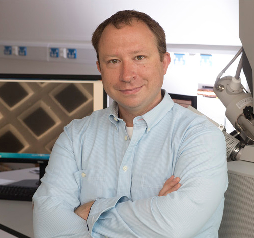
On behalf of the organising committee, I look forward to welcoming you to Sheffield, or if you are joining us virtually, online.
Stephen Muench
Chair
Format
Faraday Discussions remain amongst the only conferences to distribute the speakers’ research papers in advance, allowing the majority of each meeting to be devoted to discussion in which all delegates can participate. Following each meeting a written record of the discussion is published alongside the papers in the Faraday Discussions journal.Find out more about the Faraday Discussions in the video available.
Themes
Cryo-electron microscopy has undergone significant developments in microscope design, camera technology and data processing regimes, but there are significant challenges that remain and opportunities to explore, many of which must be tackled by the community as a whole rather than by individual groups. This meeting will bring together expertise from both these centres and the international cryo-EM community to discuss the current developments and challengesThis Faraday Discussion will be organised into the following themes:
Sample preparation in single particle cryo-EM
Sample preparation is central to electron microscopy and is currently a significant bottleneck in many experiments. There are currently a number of limitations in our fundamental understanding of the sample preparation process, in particular the way a protein sample interacts with a grid support and how this affects the sample’s integrity. The surface chemistry of these grids is poorly characterized. Discussion in this section will focus on dealing with the air/water interface, improving sample reproducibility and changing the chemistry of support grids to better facilitate sample preparation.
Pushing the limits in single particle cryo-EM
Despite the recent massive improvements in single particle cryo-EM, obtaining sub-2Å structural information is still a major challenge. There are a number of limitations that hinder our ability to obtain atomic resolution maps, including imaging conditions and data processing approaches. In this section, we will discuss what factors currently limit the resolution of most structures, and how we can quickly screen buffer conditions to optimise data quality, improve the signal-to-noise within data, accurately correct for factors such as magnification anisotropy and Ewald sphere curvature and improve current methodologies for identifying different conformational states. This session will also discuss the latest advances in phase plate technology and opportunities for further developments.
Tomographic analysis, CLEM
Many structural techniques can give atomic or near atomic level information, but many lack the ability to study proteins within a near native environment, for example within its cellular compartment. However, tomographic analysis provides both high resolution and the ability to work within the cell. With advances in labelling technologies and better stages it is now possible to conduct correlative electron microscopy (CLEM) analysis which can localise labelled proteins within the cell to high precision. This session will discuss the challenges facing this developing technology and how the community can address them. Key discussion points will be how we can build better sample stages, labelling technologies and tilt collection strategies, and if improved grid chemistry could help with sample preparation.
Map/model validation and machine learning in EM
Developments in cryo-EM are creating new opportunities within structural biology, however there are significant problems with ensuring the integrity of the field in terms of dealing with inherently low signal-to-noise images. Unfortunately, there have been recent examples in the literature of poorly refined and validated structures from cryo-EM data and with the rapid expansion of EM facilities throughout the UK and worldwide, how do we ensure that sufficient tools and training are provided to maintain high standards of model validation? Moreover, what role will the implementation of more machine learning approaches play in cryo-EM data processing and validation? In this session, we will discuss how we can ensure cryo-EM derived maps are validated, the current tools for model building and how they can be improved, and the role of ever changing data processing approaches.
Useful links
Downloads
- Programme
- Preprints - Session 1: Sample preparation in single particle cryo-EM
- Preprints - Session 1 cont: Sample preparation in single particle cryo-EM
- Preprints - Session 2: Tomographic analysis, CLEM
- Preprints - Session 3: Map/model validation and machine learning in EM
- Preprints - Session 3 cont: Map/model validation and machine learning in EM
- Preprints - Session 4: Pushing the limits in single particle cryo-EM
- Preprints - Session 4 cont: Pushing the limits in single particle cryo-EM


