

You can find details about how to access information remotely in this step-by-step guide. The guide will also help if for any reason you have difficulty accessing the content you want.
What would you like to know about this journal?
ChemComm is a Transformative Journal, and Plan S compliant
Impact factor: 4.9*
Time to first decision (all decisions): 18 days**
Time to first decision (peer reviewed only): 23 days***
Chair: Douglas Stephan
Indexed in Science Citation Index (SCI) and MEDLINE
Open access publishing options available
ChemComm is the Royal Society of Chemistry’s journal for urgent communications of outstanding significance from across the chemical sciences.
The RSC’s most cited journal, we have been one of the most trusted venues for rapid publication of short communications for more than 50 years.
ChemComm is here to support researchers throughout their careers. Whether you’re a first-time author or a senior academic you can trust us to handle your submission fairly and efficiently.
Our publication times are amongst the fastest in chemistry, and researchers typically receive a first decision on their peer-reviewed manuscript within 3 weeks of submission.
ChemComm is part of the RSC’s family of high impact general chemistry journals, alongside Chemical Science and Chemical Society Reviews.
Journal scope
ChemComm publishes urgent research which is of outstanding significance and interest to experts in the field, while also appealing to the journal’s broad chemistry readership. Our communication format is ideally suited to short, urgent studies that are of such importance that they require accelerated publication.
Our scope covers all topics in chemistry, and research at the interface of chemistry and other disciplines (such as materials science, nanoscience, physics, engineering and biology) where there is a significant novelty in the chemistry aspects.
Major topic areas covered include:
- Analytical Chemistry
- Catalysis
- Chemical Biology and medicinal chemistry
- Computational Chemistry and Machine Learning
- Energy and sustainable chemistry
- Environmental Chemistry
- Green Chemistry
- Inorganic Chemistry
- Materials Chemistry
- Nanoscience
- Organic Chemistry
- Physical Chemistry
- Polymer Chemistry
- Supramolecular Chemistry
ChemComm Emerging Investigator Lectureship Award
This award recognises emerging scientists in the early stages of their independent academic careers who have made a significant contribution to the chemical sciences.
We expect to open nominations for the 2024 lectureship in Spring.
Read about eligibility, how to nominate, and see all award winners
Meet the team
Find out who is on the editorial and advisory boards for the ChemComm journal.
Chair
Douglas Stephan, University of Toronto, Canada
Associate editors
Lutz Ackermann, University of Göttingen, Germany
Davide Bonifazi, University of Vienna, Austria
Deanna D’Alessandro, University of Sydney, Australia
Fengtao Fan, Chinese Academy of Sciences, China
Itaru Hamachi, Kyoto University, Japan
Michaele Hardie, University of Leeds, UK
Kim Jelfs, Imperial College London, UK
Chao-Jun Li, McGill University, Canada
David Lou, City University of Hong Kong, Hong Kong
Connie Lu, University of Bonn, Germany
Marinella Mazzanti, EPFL, Switzerland
Yang Tian, East China Normal University, China
Sandeep Verma, Indian Institute of Technology Kanpur, India
Brendan Abrahams, University of Melbourne, Australia
Polly Arnold, University of Edinburgh, UK
Louise Berben, University of California, Davis, USA
Akkattu T. Biju, Indian Institute of Science, Bangalore
Penny Brothers, Australian National University, Australia
Wesley Browne, University of Groningen, The Netherlands
Raffaella Buonsanti, EPFL, Switzerland
Hong Chen, Soochow University, China.
Xiao-Ming Chen, Sun Yat-Sen University, China
Arindam Chowdhury, Indian Institute of Technology Bombay, India
Derrick Clive, University of Alberta, Canada
Seth Cohen, University of California, San Diego
Marcetta Darensbourg, Texas A&M University, USA
Jyotirmayee Dash, Indian Association for the Cultivation of Science, India
Gautam R Desiraju, Indian Institute of Science, Bangalore, India
Abhishek Dey, Indian Association for the Cultivation of Science (IACS), India
Joshua Figueroa, University of California, San Diego, USA
Lutz Gade, University of Heidelberg, Germany
Sujit Ghosh, Indian Institute of Science and Education Research, India
Robert Gilliard Jr., Massachusetts Institute of Technology, USA
David González-Rodríguez, Autónoma University of Madrid, Spain
Rebecca Goss, University of St Andrews, UK
Mike Greaney, University of Manchester, UK
Shaojun Guo, Peking University, China
Michaele Hardie, University of Leeds, UK
Amanda Hargrove, Duke University, USA
Hongyan He, Institute of Process Engineering, Chinese Academy of Sciences, China
Eva Hevia, University of Bern, Switzerland
Feihe Huang, Zhejiang University, China
Todd Hudnall, Texas State University, USA
Ilich A. Ibarra Alvarado, National University of Mexico, Mexico
Ajeet Kaushik, Florida Polytechnic University
Jong Seung Kim, Korea University, Korea
Shu Kobayashi, University of Tokyo, Japan
Mi Hee Lim, Korea Advanced Institute of Science and Technology (KAIST), South Korea
Teck-Peng Loh, Nanyang Technological University, Singapore
Tien-Yau Luh, National Taiwan University, Chinese Taipei
Doug MacFarlane, Monash University, Australia
Hiromitsu Maeda, Ritsumeikan University, Japan
Silvia Marchesan, University of Trieste, Italy
Nazario Martin, Complutense University of Madrid, Spain
Alexander Miller, University of North Carolina at Chapel Hill, USA
Wonwoo Nam, Ewha Womans University, South Korea
Kenneth Ozoemena, University of the Witwatersrand, Johannesburg
Thalappil Pradeep, Indian Institute of Technology Madras, India
S Ramakrishnan, Indian Institute of Science, India
Erwin Reisner, University of Cambridge, UK
Robin Rogers, The University of Alabama, USA
Ilhyong Ryu, Osaka Metropolitan University
Paolo Samori, Université de Strasbourg, France
David Scanlon, University of Birmingham, UK
Ellen Sletten, University of California, Los Angeles, USA
David Smith, University of York, UK
Mizuki Tada, Nagoya University, Japan
Zhong-Qun Tian, Xiamen University, China
Tan Tianwei, Beijing University of Chemical Technology, China
Tomas Torres, Autonomous University of Madrid
Judy Wu, University of Houston, USA
Yi Xie, University of Science and Technology of China, China
Xianran Xing, University of Science and Technology Beijing, China
Shuli You, Shanghai Institute of Organic Chemistry, Chinese Academy of Sciences, China
Yan Yu, University of Science and Technology, China
Fan Zhang, Fudan University, China
Qiang Zhang, Tsinghua University, China
Xi Zhang, Tsinghua University, China
Wenwan Zhong, University of California, Riverside, USA
Eli Zysman-Colman, University of St. Andrews, UK
Richard Kelly, Executive Editor, 0000-0003-2380-9315
Jon Ferrier, Deputy Editor
Danny Andrews, Development Editor
Ershad Abubacker, Development Editor
Jade Holliday, Editorial Assistant
Helen Saxton, Editorial Production Manager, 0000-0002-1560-7396
Becky Webb, Senior Publishing Editor
Kirstine Anderson, Publishing Editor
Matthew Bown, Publishing Editor
Laura Cooper, Publishing Editor
Hannah Fielding, Publishing Editor
Anoushka Handa, Publishing Editor
Claire Harding, Publishing Editor
Alan Holder, Publishing Editor, ORCID 0000-0001-5228-877X
Charlie Palmer, Publishing Editor
Rosie Rothwell, Publishing Editor
Donna Smith, Publishing Editor, ORCID 0000-0002-1337-2327
Laura Smith, Publishing Editor, ORCID 0000-0002-2976-8529
Natalie Ford, Publishing Assistant
Jeanne Andres, Publisher
Journal specific guidelines
Double-anonymised peer review option
ChemComm is now offering authors the option of double-anonymised peer review. Both single- and double-anonymised peer review are now available to authors.
- Single-anonymised peer review - where reviewers are anonymous but author names and affiliations are known to reviewers (this is the traditional peer review model used on ChemComm.)
- Double-anonymised peer review - where authors and reviewers' identities are concealed from each other
Read our guidelines for authors and reviewers
Read moreGuidelines on writing titles, abstracts & table of contents information
More detailsThe title, abstract and table of contents graphic are the first parts of your manuscript that editors, referees and potential readers will see, and once published they play a major part in a researcher’s decision to read your article. Therefore it’s important that these clearly and concisely show the main findings of your research and why they are important.
Title
This should be short and straightforward to appeal to a general reader, but detailed enough to reflect the contents of the article.
- Keep it short – up to 15 words is ideal
- Use easily recognisable words and phrases that can be read quickly
- Use general terms for compounds and procedures rather than specific nomenclature, very specialised terms or non-standard abbreviations
- Avoid using terms such as “novel”. Instead say why your findings are important
- Use keywords and familiar, searchable terms – these can increase the chances of your article appearing in search results. Around 70% of our readers come via search engines
Table of contents information
This consists of a small graphic and short text which will appear in the journal contents listing
The graphic should:
- be 8 cm wide x 4 cm high
- be a clear representation of what your research is about; consider what would grab the attention as a reader scans though article listings
- include only one or two key elements; it’s much better to have a small amount of information that stands out rather than a lot of information which is too small to understand
- only include text that is large enough to read and in Arial, Times or Helvetica font
- avoid reusing figures from the article such as graphs or spectra which often, on their own, don’t convey what your research is about
Information on required formats for graphics can be found in our submission guidelines.
The text should:
- be 15–25 words
- focus only on the key findings and their importance, not the processes used; think about what would grab the attention of the reader and encourage them to read the full article
- use easily recognisable words and phrases that can be read quickly
- not repeat the information given in the title
Abstract
This is a single paragraph which summarises your research. It will help readers to decide whether your article is of interest to them.
- The length can vary, typically up to 100 words, but it should always be concise and easy to read
- It should set out the objectives of the work, the key findings and why this research is important (compared to other research in its field)
- It should emphasise (but not overstate) the significance and potential impact of your research
- Avoid including detailed information on how the research was carried out; this should be described in the main part of the manuscript
Like your title, make sure you use familiar, searchable terms and keywords
Expand for examplesTitles
Asymmetric Si-rhodamine scaffolds: rational design of pH-durable protease-activated NIR probes in vivo
From DOI 10.1039/C9CC09666C
Flexible and printable dielectric polymer composite with tunable permittivity and thermal stability
From DOI 10.1039/C9CC08648J
Carbon free silicon/polyaniline hybrid anodes with 3D conductive structures for superior lithium-ion batteries
From DOI 10.1039/C9CC09132G
Borohydride intermediates pave the way for magnesium-catalysed enantioselective ketone reduction
From DOI 10.1039/C9CC09111D
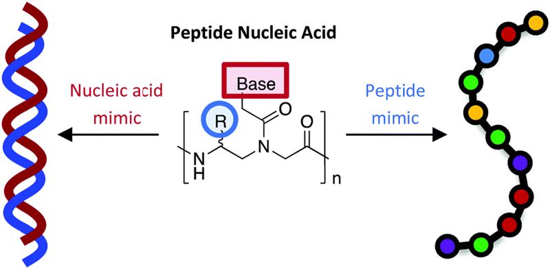
Peptide nucleic acids harness dual information codes in a single molecule
From DOI 10.1039/C9CC09905K
Table of contents entries
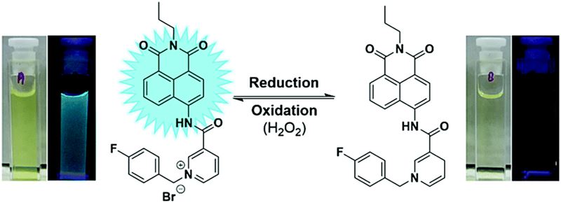
A new naphthalimide based NADH mimic that functions as a fully reversible fluorescent “on off” probe for redox state has been synthesised and evaluated.
From DOI: 10.1039/C9CC09748A
Peptide nucleic acids simultaneously encode information from nature's two fundamental biopolymers for life: nucleic acids and proteins. From DOI: 10.1039/C9CC09905K
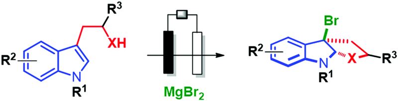
We report an efficient and environmentally friendly electrochemical approach to perform the bromo cyclization of tryptophol, tryptamine and tryptophan derivatives. From DOI 10.1039/C9CC09276E
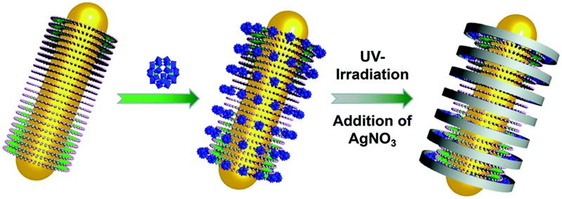
Polyoxometalates (POMs) were self-assembled on cetyltriethylammonium bromide-covered gold nanorods and formed periodic POM rings, which could be used as templates for the synthesis of Ag nano-rings. From DOI 10.1039/C9CC06968B
Abstracts
Cytochalasans are highly complex fungal metabolites which exhibit diverse biological activities. Little is known of the chemical steps involved in the construction of the tricyclic core, which consists of an octahydro-isoindole skeleton fused to a macrocyclic ring. Here, using a directed gene knockout and complementation strategy, we show that PyiF is implicated as the proposed intramolecular [4+2] Diels–Alderase required for construction of the tricyclic core of pyrichalasin H
From DOI 10.1039/C9CC09590J
A one-step process has been developed to produce a polymer coating which is hydrophobic and oleophilic, but which oil slides off and water adheres to at all tilt angles – including when vertically inclined or inverted. The polymer is transparent, and the plasma coating process is independent of substrate composition and geometry.
From DOI 10.1039/C9CC08896B
The aplyronines are a family of highly cytotoxic marine natural products with potential application in targeted cancer chemotherapy. To address the severe supply issue, function-oriented molecular editing of their macrolactone scaffold led to the design of a series of simplified aplyronine analogues. Enabled by a highly convergent aldol-based route, the total synthesis of four analogues was achieved, with a significant improvement in step economy versus previous compounds, and their cancer cell growth inhibition in the HeLa cell line was determined. The modular strategy presented offers a means for significantly shortening their chemical synthesis to facilitate the continued development of this promising class of anticancer agent.
From DOI 10.1039/C9CC09050A
Shell-isolated nanoparticle-enhanced Raman spectroscopy (SHINERS) is applied to the study of a state-of-the-art water oxidation electrocatalyst, IrOx, during oxygen evolution. The excellent sensitivity allows for in situ detection of surface intermediate species during cyclic voltammetry. Features in the Raman spectrum are correlated with the redox behaviour of the electrode, demonstrating a way to study the mechanisms of electrocatalytic water splitting using equipment available in most laboratories.
From DOI 10.1039/C9CC08284K
Conjugate addition of thiols to benzoquinones has been coupled to in situ electrochemical oxidation of the resulting hydroquinone to enable full substitution of quinone C–H bonds. The sulfonated thioether-substituted quinones exhibit high solublity and stability in aqueous solution and have redox potentials ranging from 440–750 mV vs. SHE. The electrosynthetic protocol is effective on >100 g scale.
From DOI 10.1039/C9CC08878D
Open access publishing options
ChemComm is a hybrid (transformative) journal and gives authors the choice of publishing their research either via the traditional subscription-based model or instead by choosing our gold open access option. Find out more about our Transformative Journals. which are Plan S compliant.
Gold open access
For authors who want to publish their article gold open access, ChemComm charges an article processing charge (APC) of £2,750 (+ any applicable tax). Our APC is all-inclusive and makes your article freely available online immediately, permanently, and includes your choice of Creative Commons licence (CC BY or CC BY-NC) at no extra cost. It is not a submission charge, so you only pay if your article is accepted for publication.
Learn more about publishing open access.
Read & Publish
If your institution has a Read & Publish agreement in place with the Royal Society of Chemistry, APCs for gold open access publishing in ChemComm may already be covered.
Use our journal finder to check if your institution has an open access agreement with us.
Please use your official institutional email address to submit your manuscript and check you are assigned as the corresponding author; this helps us to identify if you are eligible for Read & Publish or other APC discounts.
Traditional subscription model
Authors can also publish in ChemComm via the traditional subscription model without needing to pay an APC. Articles published via this route are available to institutions and individuals who subscribe to the journal. Our standard licence allows you to make the accepted manuscript of your article freely available after a 12-month embargo period. This is known as the green route to open access.
Readership information
ChemComm is for academic and industrial chemists in all areas of the chemical sciences.
Subscription information
ChemComm is part of RSC Gold, Core Chemistry and General Chemistry subscription packages.
Online only 2024: ISSN 1364-548X, £3,695 / $6,508
*2022 Journal Citation Reports (Clarivate Analytics, 2023)
**The median time from submission to first decision including manuscripts rejected without peer review from the previous calendar year
***The median time from submission to first decision for peer-reviewed manuscripts from the previous calendar year
ChemComm
- Email:
- Send us an email
- Email:
- Send us an email
Share
Advertisement